August 15, 2025
3 min read
First 3D Images of Human Embryo Implantation Reveal New Details of the Process
Analyzing embryo movements in uteruslike environments could offer clues to improving the success rate of in vitro fertilization
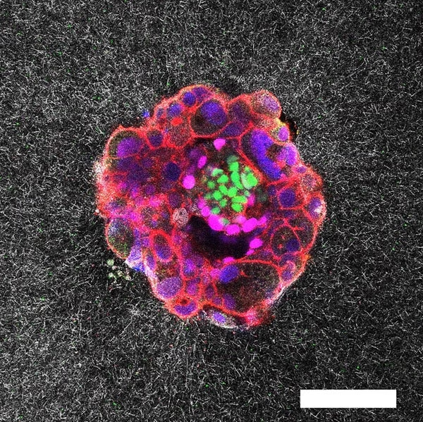
Confocal microscopy image of a nine-day-old human embryo. Specific proteins and cellular structures have been coloured in the image: OCT4 (green), which is related to embryonic stem cells; GATA6 (magenta), which is associated with early tissue formation; DAPI (blue), which marks the DNA in the nuclei; and phalloidin (red), which reveals the actin cytoskeleton. The scale bar corresponds to 100 µm.
Institute for Bioengineering of Catalonia (IBEC)
Researchers have captured the very first real-time, three-dimensional images and videos of a human embryo implanting into collagen designed to mimic uterine tissue —a key stage in reproduction. The resulting footage, which shows how embryos push and pull to anchor themselves in the uterus in vivid detail, could lead to improvements for in vitro fertilization (IVF) techniques, the scientists say.
“This will allow us to develop treatments specifically targeting implantation, which is the biggest roadblock in human reproduction,” says Samuel Ojosnegros, a bioengineer at the Barcelona Institute of Science and Technology in Spain and a co-author of the new study, which was published in Science Advances.
Five days after an embryo is fertilized artificially, fertility doctors must implant it into the body so it can continue to grow. “What happens between the transfer and the first ultrasound weeks later is a black box,” says Ojosnegros, who is also co-founder of the biotech company Serabiotics. Implantation failure is one of the main causes of infertility —up to 60 percent of miscarriages occur during this process.
On supporting science journalism
If you’re enjoying this article, consider supporting our award-winning journalism by subscribing. By purchasing a subscription you are helping to ensure the future of impactful stories about the discoveries and ideas shaping our world today.
The first successful culture of human embryos beyond implantation was demonstrated in a petri dish in a lab in 2016, but Ojosnegros and his team wanted to see what this process would look like in 3D tissue that was more similar to that of the uterus.
To do this, the team designed a special ex vivo system made of gel and collagen—a protein found in the uterine…
Click Here to Read the Full Original Article at Scientific American Content: Global…

